
Fig. 2.
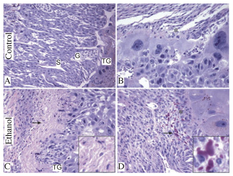
Chronic gestational exposure to ethanol alters placental morphology. Junctional zone composed of spongiotrophoblast [S], glycogen cells [G], trophoblast giant cells [TG] (A) and decidua [*] (B) is demonstrated in control placenta. Foci of necrosis in junctional zone (arrow, C) and intracellular and extracellular amorphous hyaline deposits (arrow, D) were noted within decidua basalis of placentas from ethanol-exposed dams. Insets in panels C and D show higher magnification images of necrosis and amorphous hyaline deposits, respectively (original magnification X800). (H&E stained sections, original magnification X20 [A, C] and X400 [B, D]).