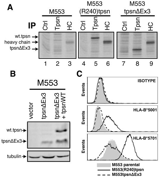
Figure 2. TpsnΔEx3 expression does not restore tapasin deficiency.
A. Tapasin deficient M553 cells or the indicated transfectants were metabolically labeled, lysed in TritonX100 containing lysis buffer and immunoprecipitations (IP) performed with control (Ctrl) antibody (L243), tapasin (Tpsn) specific antibody TO-3 or heavy chain (HC) specific antibody TP25.99. B. Lysates from M553 cells transfected with empty plasmid (vector) or tpsnEx3 were assessed for for tapasin by western blot using TO-3 antibody. Control lane shows relative position of wild type tapasin and tpsnΔEx3. C. Untransfected M553 cells (filled histograms) or stable transfectants containing wild type R240tpsn (solid line) or tpsnΔEx3 (dotted line) were examined for MHC class I expression by TT4-A20 staining (HLA-B*5701), SFR8.B6 (HLA-B*5001) staining, or isotype control staining. A representative figure of at least three independent experiments is shown.

