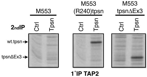
Figure 3. TpsnΔEx3 interacts with TAP.
M553 cells were treated with IFN for 16-20 hours and metabolically labeled using 35S methionine prior to lysis in digitonin containing lysis buffer. Cell lysates were subjected to immunoprecipitation (1°IP) using a TAP2 specific antibody. After elution, re-IP (2°IP) with a control antibody (Ctrl) or TO-3 (Tpsn) was performed and gels subject to autoradiography.

