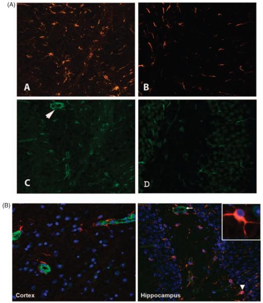
Figure 7.
C3aR antagonist treatment reduces astrogliosis, in the brains of lupus mice. Representative sections immunostained for GFAP and vimentin to determine the degree of astrocyte involvement. Sections from MRL/lpr mice treated with vehicle (A and C) or with C3aRa (B and D) are given here. Vehicle-administered mice had numerous GFAP (A) and vimentin (C)-labeled astrocytes compared with those treated with C3aRa (B and D). Photomicrographs were taken at 10X magnification with a Zeiss camera. All images are typical and representative of each group. (B) Panel A shows vimentin (green) strongly expressed around the micro-vasculature compared with GFAP (red) that was present around the periphery. However, panel B shows that both GFAP and vimentin co-localized in the astrocytes. Inset shows that GFAP is predominant in the astrocytic processes. Nuclei are stained in blue (DAPI).

