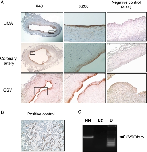
Figure 1.
HN expression in endothelial cells of human arteries and veins. (A) Immunostaining of HN (brown) at low and high magnification in normal LIMA, atherosclerotic coronary artery, and saphenous vein (GSV). Negative controls: LIMA, coronary artery, and GSV were incubated with rabbit serum instead of HN antibody. (B) HN is expressed in Leydig cells of young male testis (positive control). HN-positive cells are indicated by the brown staining at the site of the endothelium. (C) HN mRNA is expressed in HAECs. HN, RT–PCR performed on mRNA isolated from HAECs showed that HN mRNA is expressed in cultured endothelial cells. NC, negative control was performed whole reaction without DNA. D, DNA ladder.

