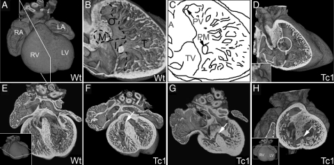
Figure 1.
E14.5 Tc1 embryos exhibit ventricular septal defects. Three-dimensional reconstructions of wild-type (A, B, and E) and Tc1 (D, F, G, and H) hearts, eroded in the planes indicated in insets. (B and C) Division of the ventricular septum (right view) into membranous (M), outlet (O), inlet (I), and trabeculated (T) regions. Many Tc1 samples have defects in the membranous region (white circle, D and inset). (E–G) Four-chambered view showing membranous VSD (white arrow, F) and muscular inlet VSD (white arrow, G; the defect has an exclusively muscular border and opens to the inlet region of the ventricular septum) in Tc1 hearts. (H) Short-axis view of a Tc1 muscular inlet VSD (white arrow, H). (LA, left atrium; LV, left ventricle; RA, right atrium; RV, right ventricle; M, membranous region; O, outlet region; I, inlet region; T, trabeculated region; A, aorta; PV, pulmonary valve; PM, base of papillary muscle; TV, septal leaflet of tricuspid valve).

