Figure 2.
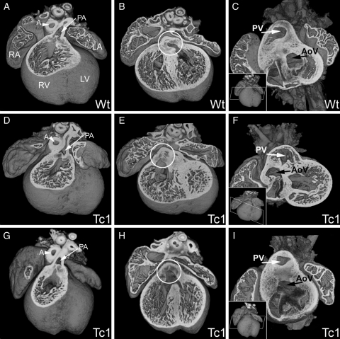
E14.5 Tc1 embryos exhibit outflow tract defects. Three-dimensional reconstructions of wild-type (A–C) and Tc1 (D–F, G–I) hearts. In all samples analysed, the pulmonary artery (PA, white arrow, A, D, and G) arose from the right ventricle (RV). In the wild-type, the aorta arises from the left ventricle (B and C), whereas in the Tc1 hearts, the position of the aortic valve (white circle, B, E, and H) is shifted rightwards. Tc1 embryos can exhibit DORV, both the pulmonary artery and the aorta arising from the right ventricle (E and F) and overriding aorta, the aortic valve (AV) sitting centrally above a VSD, resulting in subaortic interventricular communication (H and I). The models in C, F, and I have been eroded to the level of the white box indicated in the respective insets. (LA, left atrium; LV, left ventricle; RA, right atrium; RV, right ventricle; PA, pulmonary artery; A, aorta; PV, pulmonary valve; AoV, aortic valve).

