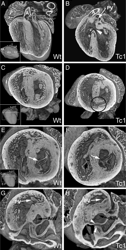
Figure 5.
E18.5 Tc1 embryos exhibit AVSD, ventricular septal, and outflow tract defects. Three-dimensional reconstructions of wild-type (A, C, E, and G) and Tc1 (B, D, F, and H) hearts eroded to the levels indicated in the insets. (B) A Tc1 membranous VSD (white arrowhead; four-chamber view) resulting in subpulmonary interventricular communication, both arterial roots arising side-by-side from the right ventricle (the Taussig–Bing malformation). (C–H) Short-axis view from apex. (D) A Tc1 muscular inlet VSD (black circle, D). The left AV valve in the Tc1 heart is trifoliate in comparison to the normal mitral valve (compare E and F, white arrows). This apparent cleft in the aortic leaflet can be traced in continuity with the two bridging leaflets (white arrowheads, H) guarding a common AV junction (see also Supplementary material online, Movie S9) (AoV, aortic valve; LA, left atrium; LV, left ventricle; RA, right atrium; RV, right ventricle; A, aorta; PV, pulmonary valve; R, right; L, left; VS, ventricular septum).

