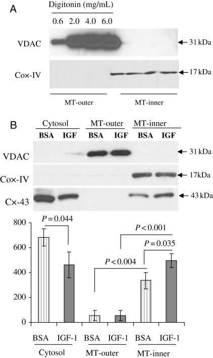
Figure 2.
Mitochondrial translocation of Cx-43 in preconditioned stem cells. (A) Western immunoblot showing purity of mitochondrial subfractionations in Sca-1+ cells using different concentrations of digitonin (ranging from 0.6 to 6 mg/mL) in the mitochondria lysis buffer. VDAC and Cox-IV were used as the mitochondrial outer and inner membrane markers, respectively. MT-outer, mitochondria outer membrane; MT-inner, mitochondria inner membrane. (B) Western immunoblot showing translocation of Cx-43 subsequent to preconditioning of Sca-1+ cells using 100 μM IGF-1 (the number of experiments performed, n = 3). BSA, bovine serum albumin.

