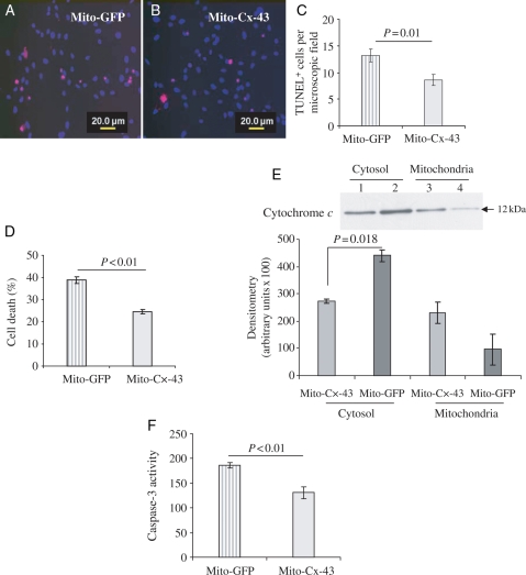
Figure 5.
Pro-survival activity of mitochondrial Cx-43 in stem cells. Sca-1+ cells were transfected with either mito-GFP-FLAG (control) or mito-Cx-43-FLAG and later were subjected to 8 h OGD. (A–C) The number of TUNEL+ cells per microscopic field (×400) was significantly reduced in Sca-1+ cells transfected with mito-Cx-43-FLAG when compared with the control (P < 0.01 vs. control cells). (D) LDH release assay showed significantly higher cell death in mito-GFP-FLAG-transfected control cells when compared with mito-Cx-43-FLAG cells (P < 0.01 vs. control). (E) Western immunoblots for cytochrome c using cell lysate samples from cytosolic fraction and mitochondrial inner membrane fraction. The cells after their transfection with the respective vectors were subjected to 8 h OGD for collection of lysate samples. The cells transfected with mito-Cx-43 showed reduced cytochrome c release from mitochondria into the cytosol (P = 0.018 vs. control; lanes 1 and 3, mito-Cx-43 transfected; lanes 2 and 4, mito-GFP transfected). Concomitantly, caspase-3 activity (F) was significantly reduced in mito-Cx-43-transfected Sca-1+ cells after 8 h OGD when compared with the control cells (P < 0.01 vs. control) (the number of experiments performed, n = 3).

