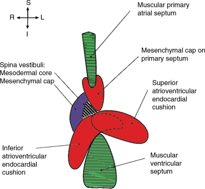
Figure 1.
Multiple tissues contribute to AV septation during development. In normal cardiac development, the AV septum is formed from myocardial septae (green), endothelially derived mesenchyme of the AV cushions and the cap of the primary atrial septum (red), and non-endothelially derived mesenchyme of the dorsal mesenchymal protrusion (aka spina vestibuli). The dorsal mesenchymal protrusion then differentiates into muscle at the AV junction (black and white stripes). Adapted from Webb et al.;13 used with permission.

