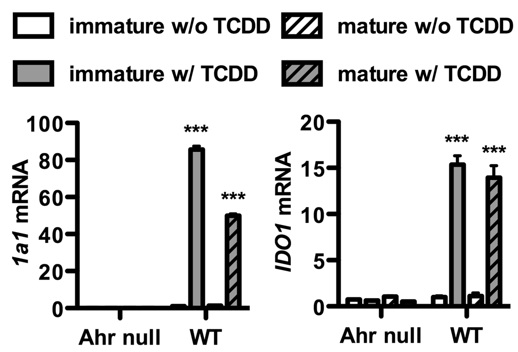
Figure 1. AHR activation in mouse BMDCs leads to IDO.
BMDCs were generated from the bone marrow of C57BL/6J wild type (WT) and AHR null mice as described in Material and Methods. Cells were harvested on day 6 as immature BMDCs or on day 7 as mature BMDCs following addition on day 6 of LPS at a dose of 50ng/ml, a concentration which itself does not cause IDO expression, confirmed in supplemental figure 1. BMDCs were cultured in the presence or absence of TCDD (10nM) added on day 0 of culture. mRNA was isolated from immature or mature BMDCs and assayed for the expression of Cyp1a1 (left panel), a marker of AHR activation, and IDO1 (right panel). Data was normalized to WT control. Post ANOVA testing comparisons are to cultures without TCDD; ***, p < 0.001. Cyp1a1 mRNA was undetectable in all AHR null PCR reactions. Each graph is representative of three independent experiments.

