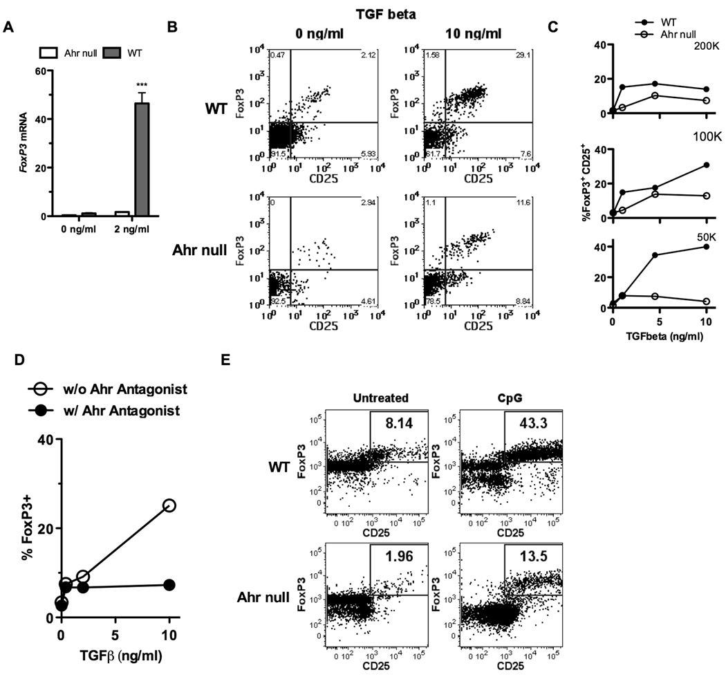
Figure 3. Presence of the AHR is necessary in T cells for optimal generation of FoxP3+ Tregs in Treg-polarizing conditions with and without cell-cell contact.
A, Naïve CD4 T cells (CD4+CD62L+ T cells) were generated by magnetic bead separation. 5×10⁵ cells/well were cultured for 5 days with anti CD3/CD28 beads in the presence of no or 2ng/ml of TGF-β. qPCR was used to test the generation of FoxP3. B, Naïve CD4 T cells (CD4+CD62L+ T cells) were generated by magnetic bead separation. 5×10⁵ cells/well were cultured for 5 days with anti CD3/CD28 beads in the presence of titrating doses of TGF-β. Flow cytometry was used to analyze for CD25 and intracellular FoxP3. C, Graphical representation of a similar experiment as B, utilizing titrating doses of TGF-β and titrating numbers of T cells per well. D, Similar to C, except some naïve cells were exposed to a soluble AHR antagonist. E, Naïve CD4+ CD25− T cells were isolated from B6 WT and AHR-null mice and co-cultured with pDCs isolated from BALB/c mice using the Miltenyi mouse pDC isolation kit at a ratio of 20 to 1. CpG was added at the start of culture in some experiments. On day 5, cells were harvested and subjected to flow cytometric analysis. Percentages are the fraction of gated live CD4+ cells that were FoxP3/CD25 double positive. The figures are representative of 3 independent experiments.

