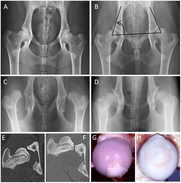
Figure 2. Radiographs and computed tomography (CT) images of the canine hip joint.
A: Unaffected hips. B: Moderate hip dysplasia. The arrow indicates the included angle which is the Norberg angle. C: Severely affected hip dysplasia with luxation. D: Severe secondary osteoarthritis as a result of previous hip dysplasia. E: A CT image of a hip with moderate hip dysplasia illustrating the subluxation and impingement of the femoral head on the lateral acetabular rim when the dog is imaged with its hips in a weight bearing position. F: A CT image of a dog with severe hip dysplasia characterized by complete luxation. G: The gross appearance of a femoral head from a hip joint moderately affected with HD and early secondary OA with articular cartilage fibrillation in the perifoveal area and hypertrophy of the teres ligament. H: A femoral head from a severely affected hip with full thickness articular cartilage erosion and loss of the teres ligament due to mechanical abrasion and enzymatic degradation. These affected hips are painful.

