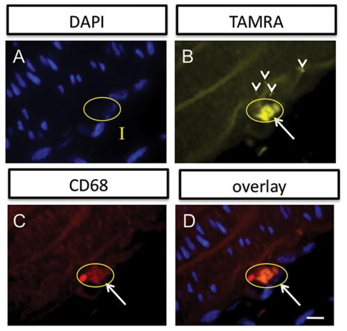
Figure 4. Immunohistochemistry demonstrates that the multimodal probes accumulate in macrophages found in the intimal layer (I) of the injured vessel wall.
(A) Cell nuclei stained with DAPI (100X). A single cell is circled in yellow that will be referenced in the other panels. (B) The TAMRA signal from the multimodal agent is found in several locations in the intima. It can be seen in places to be in a punctate patern (arrowheads), suggesting internalization. A pronounced accumulation is shown for the circled cell. (C) Anti-CD68 monoclonal antibody staining also highlights a region in the intima (circled cell at white arrow). (D) Overlay of the three channels, A-C, demonstrates that the TAMRA signal colocalizes with the CD68 positive cell with the nucleus indicated in A, supporting macrophage labeling by the probes. Scale bar = 25 microns.

