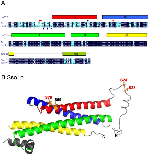
Figure 1. A schematic diagram illustrating the Sso1p and Sso2p homology and the domain structure of Sso1p.
A) Habc, H3 SNARE motif, and transmembrane domain (TMD) are indicated. Serine 23, serine 24, serine 79 in Sso1p (red arrows) and serine 31 and serine 34 in Sso2p (blue arrows) indicate the identified in vivo phosphorylation sites [22], [23]. Additional amino acids mutagenized (Serine 59 in Sso1p and Threonine 28 in Sso2p) are indicated by black arrows. B) The three dimensional structure of Sso1p (PDB 1FIO, [8]) with an added random N-terminal peptide for amino acids 1–30. For the phosphoamino acids the side chains are shown. Phosphorylation sites identified by mass spectrometry or in vivo labeling are indicated by red colour. The additional amino acid mutagenized (Serine 59) is indicated by black colour.

