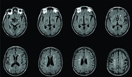
Figures 1.
T2 FLAIR image acquired from a Phillips 1.5 scanner. Case 1 is a 43-year old woman with septic shock. Magnetic resonance imaging (MRI) was performed to evaluate delirium without focal neurologic findings following a brain computed tomograpy scan. Images are axial slices, from the top right to left and then bottom left to right, starting through the temporal lobes progressing higher in the brain through the frontal and parietal lobes. The patient did not have baseline cognitive deficits (IQCODE score <3.3). Patient exhibited no obvious subarachnoid hemorrhage, ischemic stroke, brain abscess, or intracranial neoplasm on the usual care clinical MRI. Periventricular patchy white matter lesions are seen in the lower three images. The three-month follow up neuropsychological evaluation showed severe impairment in executive function (memory and attention were not assessed as the patient was too weak to perform all tests).

