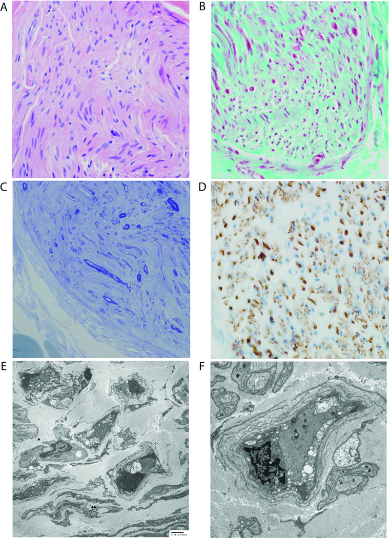
Figure 3.
Sural nerve biopsy (Case 2) stained with (A) H&E (400×), (B) trichrome (400×), and (C) toluidine blue (400×) and (D) immunohistochemistry for neurofilament protein (400×) show severe hypomyelination with intact axons. (E, F) Transmission electron microscopy reveals a dramatic reduction in the myelination of fibers and very thin myelin sheaths around infrequent fibers. Acute myelin breakdown and well-developed onion-bulb formation are focally identified.

