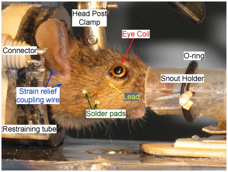
Fig. 2.

Testing setup. Lower power photo of mouse in restraining tube with homemade snout holder to deliver gas anesthesia (not shown) while coil is placed on the eye. The O-ring holds the nose in the modified syringe by securing the paper clip (barely visible) that slides in the slots. The strain relief-connecting wire leads from the solder pads to the connector and from there to the headstage. The curvature of the eye coil can be seen in this photo compared to the original in Fig. 1.
