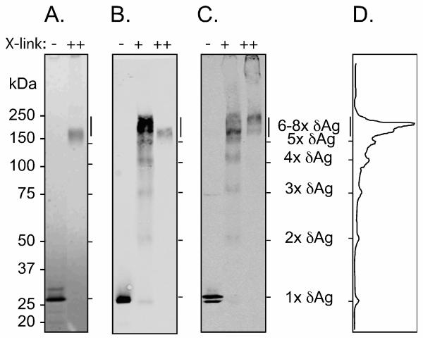
Fig. 4.
Electrophoretic mobility of purified δAg and of δAg assembled into virus-like particles. In panels A and B purified δAg, at 2 μM was treated without or with glutaraldehyde cross-linking, 0.01 or 0.1%, as indicated by + and ++ respectively. This was followed by SDS denaturation and electrophoresis on a gel of 4-12% polyacrylamide. In panel A, total protein was detected by SimplyBlue staining and in panel B, δAg was detected by immunoblot using specific antibody. In panel C, virus-like particles containing δAg, without and with prior cross-linking were examined as in panel B. The minor band migrating faster than the monomer might be a proteolytic fragment. At left are indicated MW markers. Panel D is the quantitation of panel B, lane 2. Indicated an interpretation of detected bands as multimers of δAg.

