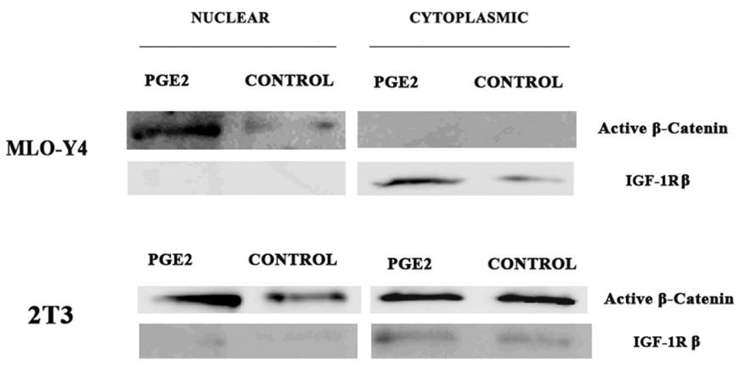
Figure 5.
Immunoblot detection of cytoplasmic and nuclear β–catenin. MLO-Y4 and 2T3 cells were grown on collagen coated dishes under static conditions and treated with PGE2 (1000 pg/ml) or vehicle (media) for 2 hours. Cells were harvested and cytoplasmic and nuclear extracts were prepared and immunoblotting performed with an antibody against the active form of β–catenin (Cell Signaling Technologies). To test the relative purity of the preparations the same blots were probed with an antibody against IGF-1R, which is a cytoplasmic protein.

