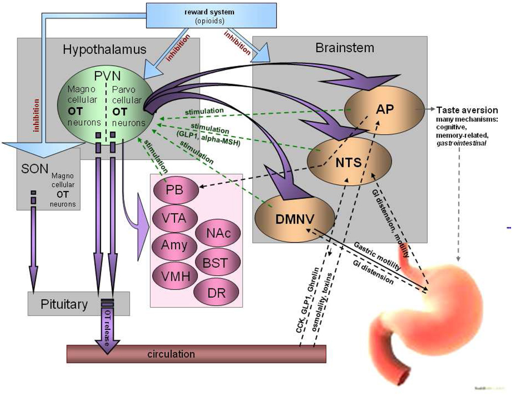
Fig. 1. Topography of central OT pathways involved in food intake regulation with special emphasis on functional significance of the circuits.
OT neurons project from parvocellular PVN neurons in the hypothalamus to brainstem sites known to regulate feeding (NTS, AP, DMNV) and to the pituitary, where OT is released to the general circulation. While the brainstem-hypothalamus pathways have been extensively studied in relation to OT’s involvement in anorexigenic responses stemming from peripheral parameters, such as GI tract distension, osmolality of the blood, etc., many other feeding-related sites that contain OT terminals or OT receptors have not been comprehensively evaluated in relation to anorexigenic action of OT. These include areas involved in reward (ventral tegmental area, VTA; nucleus accumbens, NAc; bed nucleus of the stria terminalis, BST), affect (dorsal raphe nucleus, DR), energy homeostasis (ventromedial hypothalamic nucleus, VMH) and stress (amygdala, Amy; and parabrachial nucleus; PB) (Buijs et al. , 1983, De Vries and Buijs, 1983, Kirchgessner, Sclafani, 1988, Olson et al. , 1993).

