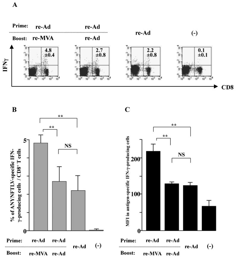
Fig. 2.

The intracellular IFN-γ staining of the ANYNFTLV, a peptide derived from Trypanosoma cruzi surface antigen-specific CD8+ T cells induced by the use of recombinant adenovirus (re-Ad) and/or recombinant vaccinia virus (re-MVA) for vaccination. (A) The representative data of intracellular IFN-γ staining of ANYNFTLV-specific CD8+ T cells derived from the re-Ad and / or re-MVA-immunized mice. B6 mice were first primed with 5 × 10⁷ plaque forming units (PFU) of re-Ad and then were given a booster immunization 14 days later with 5 × 10⁷ PFU of either re-Ad or re-MVA. The single re-Ad immunized and non-immunized mice were included as controls. The mice were sacrificed 12 days after the last immunization, and spleens were removed. The freshly-isolated splenocytes were subjected to the FACS analyses for the intracellular IFN-γ+ staining in response to the ANYNFTLV-pulsed EL-4 cells. The cell culture condition for the assays was described in the Materials and methods. The number of IFN-γ+ cells that appeared against peptide-unpulsed EL-4 was subtracted from the number of IFN-γ+ cells that appeared against peptide-pulsed EL-4. (B) The percentage of IFN-γ+ cells among total CD8+ T cells was calculated. Data represent the mean ± S.D. of three mice in each group. **, P < 0.01 determined by the Student's t-test. “NS” means “not statistically significant”. (C) The mean fluorescence intensity (MFI) of ANYNFTLV-responding IFN-γ+ cells was calibrated by gating cells which were both IFN-γ-positive and CD8-positive. Data represent the mean ± S.D. of three mice in each group. **, P < 0.01 determined by the Student's t-test. “NS” means “not statistically significant”.
