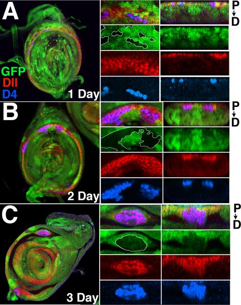
Figure 6.
D4/lacZ activation in Antp- clones of increasing age in second leg discs. Antp- clones are marked by the loss of GFP (green), and all discs are stained for Dll (red) and D4/lacZ (blue). Left-hand panels show merged images of the entire disc. The central panels show an enlarged region, with the merged image at top. The right hand panels show cross sections at the same level as the central panels. Distal extension of clones from the ring is seen as downward extension in these cross sections. (A) 1 day old Antp- clones. Note in central panels that Antp- clones activate D4/lacZ in the distal part of the Dll ring, but not in the proximal portion. No distal extension of the D4/lacZ-expressing clones has taken place. (B) By day two, D4/lacZ-expressing clones are beginning to round up and distort the ring (central panels). Slight distal extension of these clones has occurred (right panels). (C) By day three, rounding up of D4/lacZ-expressing clones is advanced (middle panels), and significant distal extension is seen (right panels).

