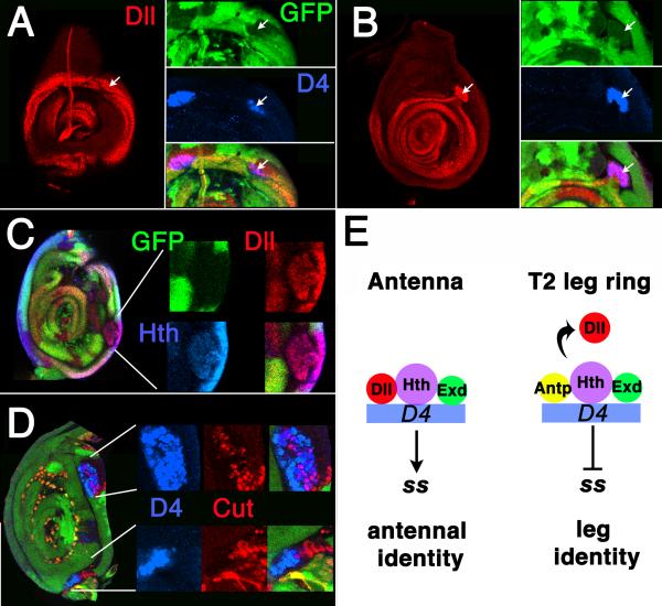
Figure 7.
In all panels, Antp- clones are marked by the loss of GFP (green). (A-B) A D4/lacZ-expressing Antp- clone in a second leg disc that extends from the proximal ring to the central domain of Dll expression. (A) Proximal focal plane, showing the Dll ring. Note activation of D4/lacZ in a clone overlapping the ring (arrows). (B) Distal focal plane, showing that the same transformed clone (arrows) connects to the distal domain of Dll expression. (C) A transformed Antp- clone in the second leg stained for Hth and Dll expression. Part of the clone has rounded up, but remains connected to the ring by a narrow isthmus. In some transformed clones, the connection is much narrower and thread-like. (D) Second leg disc containing Antp- clones stained for Cut and D4/lacZ expression. Note two rounded-up clones in which both Cut and D4/lacZ are expressed. Although Cut and D4/lacZ are coexpressed in many cells in the upper clone, Cut is expressed adjacent to a D4/lacZ expressing region in the bottom clone. (E) Model summarizing the control of D4 in the antenna and leg. Left: In the antenna, D4 is activated by binding of a Dll/Hth/Exd complex. Right: In the proximal ring of the leg, Antp displaces Dll and prevents activation of the enhancer.

