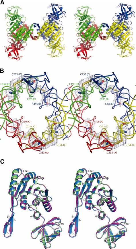
Figure 1.
Overall organization of PabTrmI (Stereoviews). (A) PabTrmI tetramer structure in complex with SAH. Subunits A and B, colored red and green, respectively, form one tight dimer and subunits C and D, colored yellow and blue, respectively, form another one. SAH is shown in blue sticks. (B) Detail of the four inter-monomer disulfide bonds that stabilize the PabTrmI tetramer. A 2 Fobs–Fcalc electron density map contoured at the level of one standard deviation is superimposed on the structure. (C) Superposition of one PabTrmI monomer from Crystal form I (in magenta), Crystal Form II (in blue) and Crystal Form III (in green).

