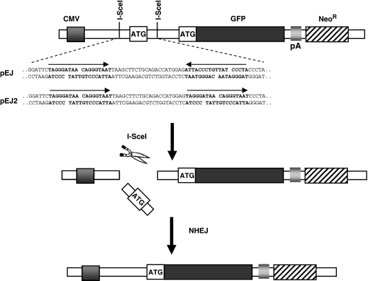
Figure 1.
Structure of the NHEJ reporter constructs pEJ and pEJ2. GFP translation is prevented by insertion of an artificial ATG (start codon) into the 5′-untranslated region between CMV promoter and the ORF. The artificial ATG is not in frame with the original ATG (17). Two repeat I-SceI recognition sequences (bold characters) flank the artificial ATG either in direct (pEJ2) or in inverted orientation (pEJ). Simultaneous cleavage of both I-SceI sites leads to pop out of the artificial ATG (middle) leaving either non-compatible ends (pEJ) or fully compatible ends (pEJ2). Repair of the I-SceI-induced DSB by NHEJ restores GFP translation leading to green fluorescence.

