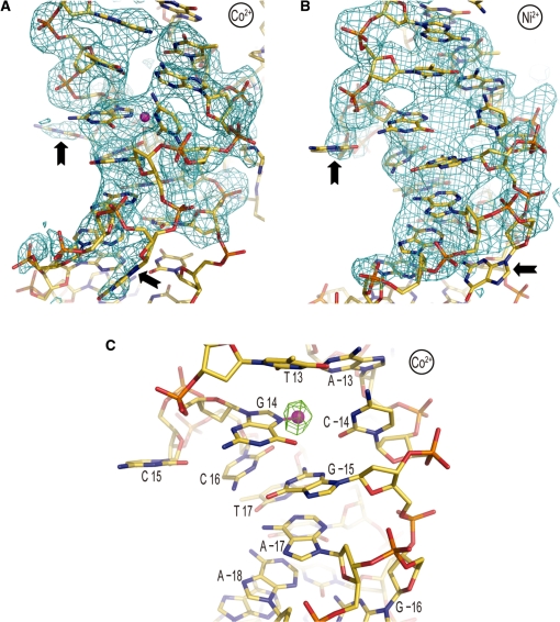
Figure 2.
Co2+ or Ni2+ binding promotes base expulsion from the double helical stack at the 1.5-turn location. (A and B) Co2+-NCP (A) and Ni2+-NCP (B) display flipping out of cytosine 15 and guanine −16 (arrows). An Fo−Fc electron density map (2.5σ), in which ATGCCTT/AAGGCAT nucleotides ±12 to ±18 were omitted from the respective models, is displayed superimposed on the structures. (C) Heavy divalent metal binding appears to directly promote the double helix deformation. Co2+ (magenta sphere) is observed to coordinate to the N7 atom of guanine 14, which is situated in the ‘minor groove’ by virtue of assuming the syn glycosidic conformation. An anomalous difference electron density map (3.5σ) is shown superimposed on the Co2+-NCP model.

