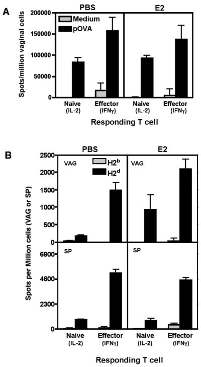
FIGURE 2. Vaginal APC from estradiol-treated mice are functional and can stimulate CD8+ T cell responses ex vivo.
A, Vaginal cells were extracted from PBS or E2 treated mice and pulsed with 10μg/ml of pSIINFEKL (pOVA) peptide or medium alone at 37°C for 1.5 hours. Pulsed cells were analyzed in Reverse Elispot assays containing either sorted naïve (IL-2) or in vitro-activated effector (IFNγ) OT-I cells. Graph shows mean±SEM of the frequency of vaginal APC inducing IFNγ or IL-2 secretion. Graph representative of three experiments. B, Cells were extracted from vaginal tissues (VAG) or spleens (SP) of PBS or E2 treated mice, and analyzed in Reverse Elispot assays using naïve anti-H2d 2C cells (IL-2) or in vitro activated effector anti-H2d 2C cells (IFNγ). Graphs show mean±SEM of the frequency of APC. N=3 mice per group. Graph representative of three independent experiments. Non-significant groups had a p-value greater than 0.05.

