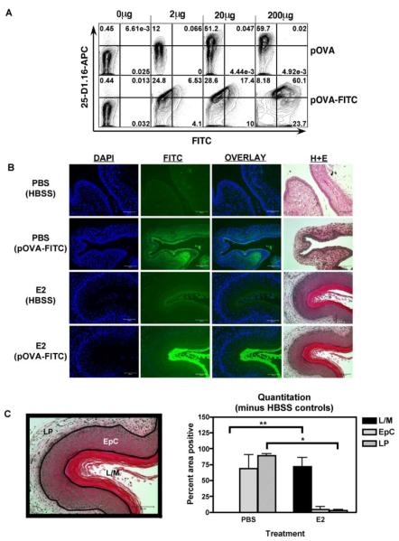
FIGURE 4. Estradiol inhibited penetration of pOVA-FITC into the vaginal wall.
A, B6 female splenocytes were pulsed with FITC-labeled or unlabeled pSIINFEKL peptide at 0μg, 2μg, 20μg or 200μg/ml in cell culture medium for 1.5hrs, stained with 25-D1.16 antibody, then analyzed by flow cytometry. B, PBS or E2 treated mice were immunized IVAG with pSIINFEKL-FITC peptide at 20μg per mouse or HBSS alone. Twelve hours after immunization vaginal tissues were fixed, embedded, and sectioned. Sequential sections were stained with the nuclear dye DAPI or with haematoxylin and eosin. Exposure times were 0.5 seconds for DAPI and 15 seconds for FITC. Representative image of an experiment repeated twice. C, The percent of the area staining positive for pSIINFEKL-FITC binding (mean±SEM) is shown in the graph, and an example of boundary assignments is shown on the left. N=3 mice per group; * and ** indicates p<0.05; Lumen/mucus (L/M); Epithelial layer (EpC); Lamina propria (LP).

