
Figure 1.
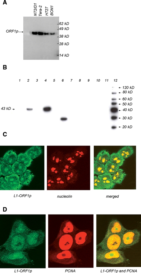
Western blot and immunofluorescence experiments using anti-L1-ORF1p sera. (A) and (B) Protein extracts were prepared from human cell lines or from normal or cancerous human tissue prior to separation by SDS-PAGE and Western blotting. In (A), extracts were from cell lines Tera-2, NTera2/D1,NCI-H727, and BON1. In (B), lanes 1 and 2 are extracts from normal adjacent tissue and breast tumor from patient “A”; lanes 3 and 4 are from normal adjacent tissue and breast tumor from patient “B”; lanes 5-10 are from normal uterine, spleen, ileal, lung, colon, and pancreatic tissue, respectively; lane 11 contains no protein extract; and lane 12 contains molecular weight markers. (C) and (D) Immunofluorescence experiments using lung cell line H460 (C) or colon cell line H1299 (D). In (C), nucleolin is used to image nucleoli, and L1-ORF1p appears to locate in the cytoplasm and nucleoli of the H460 cell line. In (D), PCNA antisera were used as these can stain nuclei but not nucleoli in HCT116 (data not shown), and the L1-ORF1 protein appears to localize in both nuclei and nucleoli but not in the cytoplasm of this cell line.