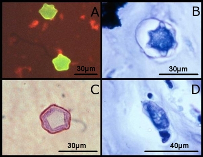
FIG. 6.
Fixed, stained sections from corneal scrapings showing cysts and a trophozoite of Acanthamoeba. (Courtesy of Paul Badenoch.) (A) Acanthamoeba cysts stained with calcofluor white. (B) Acanthamoeba cyst stained with a Giemsa stain. (C) Acanthamoeba cyst stained with a Gram stain. (D) Acanthamoeba trophozoite stained with a Giemsa stain.

