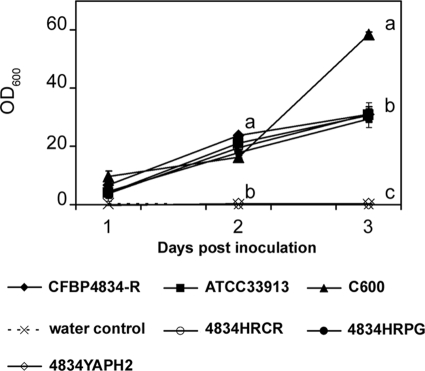
FIG. 3.
Kinetics of the adhesion capacities of bacterial strains on a polypropylene surface. X. citri pv. phaseoli var. fuscans strains CFBP4834, 4834HRCR, 4834HRPG, and 4834YAPH2; X. campestris pv. campestris ATCC 33913; and E. coli C600 were cultured during 3 days at 28°C under static conditions in polypropylene microtiter plates from an inoculum at 5 × 10⁵ CFU ml−1. Crystal violet-stained surface-attached cells were quantified by solubilizing the dye absorbed by adherent cells, after the removal of suspensions, in ethanol and determining the optical density at 600 nm. Means and SEM were calculated for data from two independent experiments, each containing three replicates per treatment and per sampling date. For a given sampling date different letters refer to significantly (P < 0.05) different values based on a Mann-Whitney U test.

