FIG. 1.
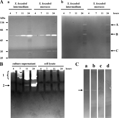
Zymographic detection of Xenorhabdus proteases in culture. (A and B) Enzyme activities produced by X. kozodoii Morocco and Intermedium strains were monitored with zymography coupled to SDS-PAGE (A) and zymography coupled to native PAGE (B). (A) The activities were tested with both casein (a) and gelatin (b) as the substrate. The positions of enzyme activity bands A, B, and C are shown to the right of the gels. (B) Protease production of X. kozodoii Morocco strain is shown using casein as the substrate. The positions of enzyme activity bands 1 and 2 are shown to the left of the gel. (C) Effects of inhibitors of catalytic serine (PMSF) and metal ion (EDTA and 1,10-phenanthroline) are shown on the activity of ∼5.0 pmol of protease B (arrow), purified from X. kozodoii Morocco strain. The gel slices are as follows: a, enzyme not treated (control); b, PMSF-treated enzyme; c, EDTA-treated enzyme; d, 1,10-phenanthroline-treated enzyme. The concentration of inhibitors during both sample treatment and gel slice incubation was 5.0 mM.

