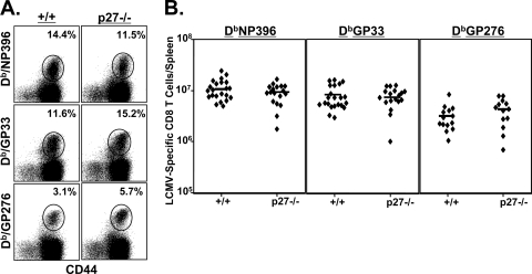
FIG. 1.
Primary CD8 T-cell response to LCMV in p27Kip1-deficient mice. Wild-type and −/− mice were infected with LCMV, and LCMV-specific CD8 T cells were quantitated in the spleens at day 8 p.i. by staining with MHC-I tetramers, anti-CD8, and anti-CD44. The dot plots in panel A are gated on the total CD8 T-cell population, and the numbers indicate percentages of LCMV-specific CD8 T cells among the total CD8 T-cell population. In panel B, each symbol represents an individual mouse. The data in panel A are from one of six independent experiments, and the data in panel B are pooled from six independent experiments.

