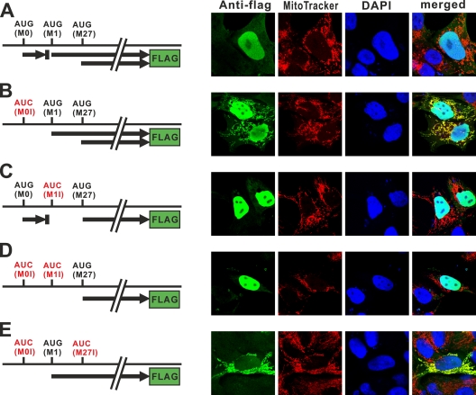
FIG. 4.
Subcellular localization of Flag-tagged mouse RNase H1 in HOS cells. HOS cells transfected with plasmids (A to E) producing Flag-tagged mouse RNase H1 were analyzed by confocal microscopy. Plasmids used in panels B to E contain AUG-to-AUC mutations, as indicated by the red color. Thick arrows represent ORFs. Cells were incubated with primary antibodies to Flag tag and secondary antibodies emitting green. Mitochondria and nuclei were stained with MitoTracker (red) and DAPI (blue), respectively. We also examined constructs in which all three AUG codons were changed to AUG, and as expected, we observed no Flag signal.

