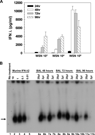
FIG. 2.
Dose dependence of IFN-λ induced by WSN virus infection. (A) Wild-type BALB/c mice (n = 4) were infected i.n. with 102, 104, or 106 PFU of influenza A/WSN/33, and BAL samples from infected animals were harvested 24 (black bars), 48 (white bars), 72 (gray bars), or 96 (hatched bars) hours postinfection. IFN-λ protein levels were determined by ELISA. Data represent the means and standard errors of the means. (B) Chinese hamster cells expressing a chimeric human IFN-λ receptor complex were left untreated (lane 1), treated with recombinant murine IFN-λ2 at 10 ng/150 μl, 1 ng/150 μl, 0.1 ng/150 μl, and 0.01 ng/150 μl (lanes 2 to 5, respectively), or incubated with BAL samples from BALB/c mice infected i.n. with 106 PFU of WSN. Either 20 μl (a) or 2 μl (b) of BAL fluid was added to the samples, for a total volume of 150 μl. Shown are assay results from individual animals at 48 h (lanes 6 and 7), 72 h (lanes 8 and 9), and 96 h (lanes 10 and 11) postinfection. After a 15-min incubation with the sample, the indicator cells were lysed, and nuclear extracts were assayed by EMSA for STAT1 activation. Recombinant murine IFN-λ2 of a known concentration was used as a standard (lanes 2, 3, 4, and 5). The arrow denotes STAT1:STAT1 homodimers. No signal was present in BAL fluid harvested from uninfected mice (data not shown).

