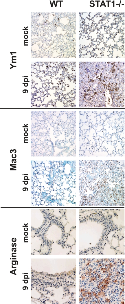
FIG. 8.
Detection of AAM markers in lung tissue of infected STAT1−/− mice by the use of immunohistochemistry. Lung sections were stained for the AAM markers arginase I, Ym1, and Mac3 with a hematoxylin counterstain. At 9 dpi, prominent staining is observed in STAT1−/− mice infected with rMA15 compared with uninfected animals and WT animals infected with rMA15. Magnification, ×40.

