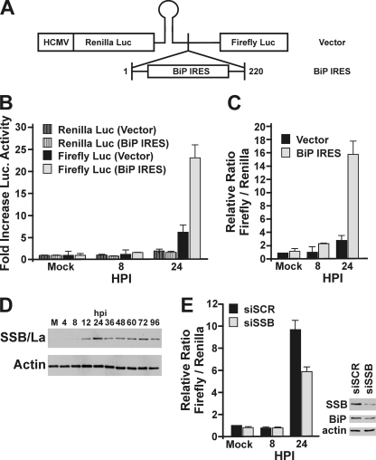
FIG. 6.
HCMV activates translation from the BiP IRES through induction of SSB/La. (A) Schematic of dual-luciferase plasmid generated by inserting the 221-nucleotide 5′ UTR of BiP (BiP IRES) into pDL-N (vector). (B and C) HFs were electroporated with the control dual-luciferase plasmid or the dual-luciferase plasmid containing the BiP IRES upstream of the firefly luciferase. Twenty-four hours postelectroporation, the cells were mock infected or HCMV infected. Protein lysates were harvested at 8 and 24 hpi. Results are graphed as fold increase in luciferase activity relative to the level in mock-infected samples (B) or as the relative ratio of firefly to Renilla luciferase for each sample, where the ratio of the mock-infected vector control was set at 1 (C). (D) Proteins from mock-infected (M) and HCMV-infected HFs were harvested at the indicated times postinfection. Western analysis was performed using antibodies against SSB/La autoantigen and actin (loading control). (E) Mock- and HCMV-infected HFs were electroporated with the dual-luciferase BiP IRES plasmid and siRNA against SSB/La (siSSB) or a control scrambled sequence (siSCR). Protein was harvested for luciferase and Western analysis at the indicated times postinfection. Results are graphed as the relative ratio of firefly to Renilla luciferase for each sample, where level of the mock-infected siSCR samples was set at 1. Western analysis was done to detect the levels of SSB/La, BiP, and actin.

