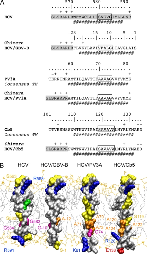
FIG. 3.
Comparison of the TMDs of HCV NS5B, GBV-B NS5B, PV3A, and Cb5. (A) HCV/PV3A and HCV/Cb5 TMD chimeras were designed in analogy to the HCV/GBV-B construct, as detailed in the legend to Fig. 2A. Amino acids are numbered with respect to PV3A and Cb5 proteins. For clarity, the ClustalW sequence comparisons are not reported. (B) Amino acid surface representations. Amino acid color coding and labeling are as described in the legend to Fig. 2E. The structure models of the HCV/PV3A and HCV/Cb5 TMDs were constructed as described in the legend to Fig. 2E. In the case of the HCV/PV3A chimeric protein, segment 16 to 44 of the Pf1 major coat protein (PDB entry 2KLV) and segment 21 to 31 of photosystem I subunit PSAX (PDB entry 1JB0) served as templates. The HCV/Cb5 three-dimensional model is based on comparison with segment 198 to 228 of aquaporin (PDB entry 3LLQ). The N termini of the chimeras were positioned at the same location with respect to the membrane in order to simplify comparisons.

