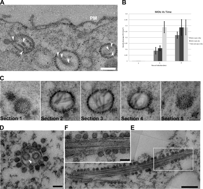
FIG. 1.
CPV-II distribution and morphology in SFV-infected BHK-21 cells. (A) CPV-II can be found close to the plasma membrane at the late stage of SFV infection. Tubular structures were well preserved in CPV-II and attached by gold particles (white arrowheads) that registered the location of anti-E2 antibodies. The gold particle was identified by its unique profile, the darkest-contrast and sharply defined edge. A few gold particles located on the plasma membrane are not marked. (B) The plot shows the CPV-II density in the BHK-21 cells that were infected with SFV at an MOI of 2 for 30 min, an MOI of 2 for 60 min, and an MOI of 200 for 30 min. The number of type II CPVs was counted after fixation of the cells at 3, 5, and 8 h postinfection and normalized relative to the measured specimen area. The average CPV-II density (number per μm2) ± standard deviation (SD) at each time point is shown. There was no CPV-II observed in mock-infected BHK-21 cells; thus the mock infection data were omitted from the graph. ads, adsorption. (C) Five consecutive sections (95 nm thick) of the same type II CPV, showing the CPV from the top to bottom. (D) A view of a CPV-II enclosed tubular structure (white arrowhead), attached by dense circular particles on the exterior surface. The two tubules within CPV-II are approximately 50 nm in diameter. (E) A longitudinal section of CPV-II shows its dimension (exceeding 1 μm). (F) A zoomed-in image of the area enclosed in a white box in panel E, showing the dense particles attached to the external membrane layer of CPV-II and the tubules. Scale bars, 200 nm (panels A, C, and E) and 50 nm (panels D and F).

