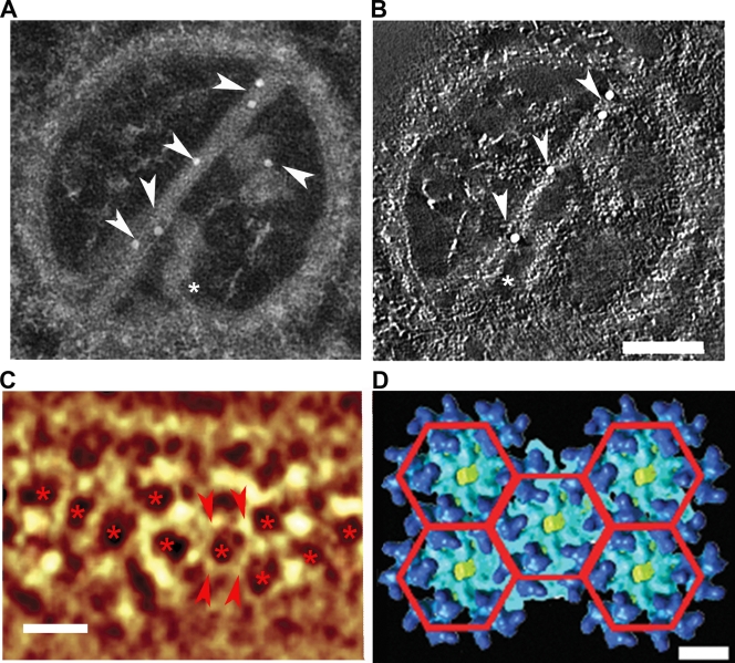
FIG. 2.
The composition of a CPV-II tubular structure. (A) The gold particles (arrowheads) were attached to the tubular structures of the tomogram of CPV-II during immunolabeling with anti-E2 antibody. Additional short tubules were observed lying vertically to the long tubules, and one of them (asterisk) appears to make contact with the CPV-II membrane. (B) An orthoslice (0.67 nm in thickness) was taken from the tomogram of the same CPV-II as in panel A, showing two tubules that appeared to have helical turns. The gold particle on the short tube is omitted from the slice, as it was positioned on the other side of the section (see also Video S1 in the supplemental material). Note that the gold labeling was performed on both sides, and it resulted in an uneven distribution of gold particles in the z dimension. Scale bar, 100 nm. (C) Volume rendering of CPV-II tubule tomogram shows the hexagonal array (asterisks) of the density nodes. Scale bar, 15 nm. The node-to-node distance (between the pair of arrowheads) is approximately 7 nm. (D) A planar array of five adjacent hexagonal rings of SFV glycoprotein trimers indicates that the trimer-to-trimer distance is 7.2 nm. Scale bar, 7 nm.

