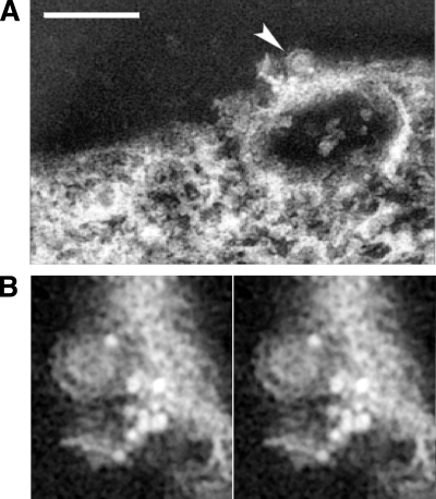
FIG. 3.
Arrangement of viral glycoproteins at the site of budding on the plasma membrane. (A) A volume projection of SFV budding site reveals that the budding virus (arrowhead) was in proximity to the gold particles that registered the location of the E2 glycoprotein on the plasma membrane. Scale bar, 200 nm. (B) The stereo pair of projection views above plasma membrane illustrates the budding region around an exiting virion (arrowhead in panel A). This close-up view shows the exterior surface of the plasma membrane with detailed arrangement of an associated group of gold markers, with the majority of them observed with an average spacing of ∼7 nm. The conjugated gold particles are located on the plasma membrane with the brightest density as a result of the high-angle electron scattering in electron microscopic imaging.

