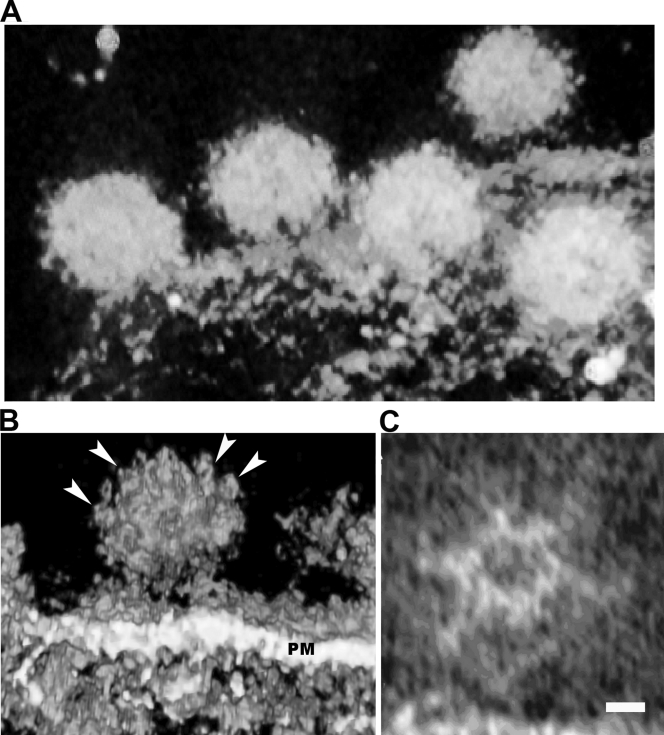
FIG. 4.
Array of glycoproteins on budding SFV. (A) A volume rendering of the tomogram on the budding viruses on the plasma membrane of a cell infected with SFV. Several budding virions (∼70 nm in diameter) were captured before scission from plasma membrane. (B) Surface rendering of this tomogram showing the interstices of the spikes on the outer envelope of the virion. A budding virion clearly shows the hexagonal array of the glycoproteins (arrowheads), where the node-to-node distance is measured as 7 nm. PM, plasma membrane. (C) A slice section from the tomogram of the same budding virus demonstrates clearly the hexagonal density, corresponding well to the glycoprotein network in the cryo-EM density map of isolated virus. Scale bar, 5 nm.

