FIG. 2.
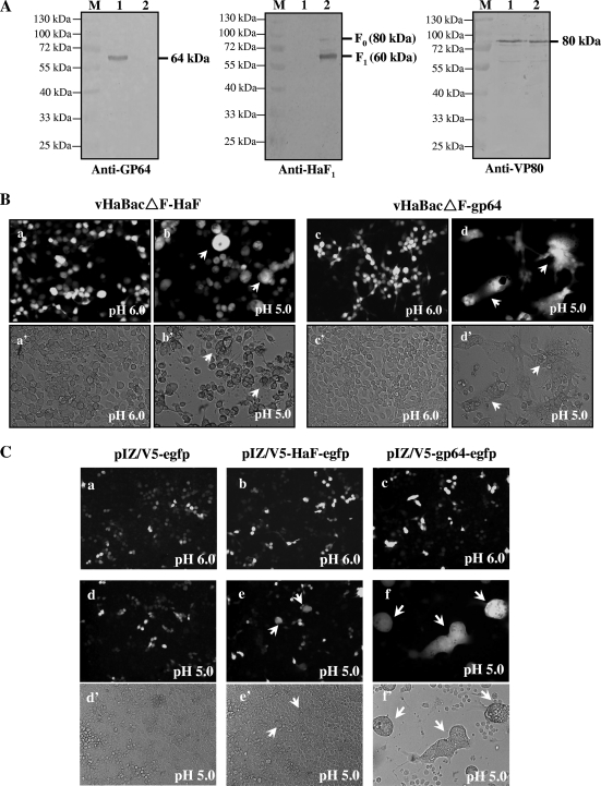
Detection of GP64 expression and fusogenicity. (A) Western blot analyses of envelope protein in recombinant BV. Virions were harvested and purified from the supernatants of infected HzAM1 cells at 6 days p.i., and the proteins were separated by SDS-PAGE. The blots were probed with antibodies against GP64 (left), HaF1 (middle), and VP80 (right). Lanes: 1, HaBacΔF-gp64 BV; 2, vHaBacΔF -HaF BV; M, the protein marker with indicated molecular mass. (B) Syncytium formation assay of HzAM1 cells infected by recombinant viruses. HzAM1 cells were infected with vHaBacΔF-HaF or vHaBacΔF-gp64 at 5 TCID50 units/cell. At 48 h p.i., cells were incubated with Grace's insect medium at pH 6.0 (a and c) or 5.0 (b and d) for 5 min. Syncytium formation was examined by fluorescence microscopy 24 h after dropping the pH. Images in panels a to d were taken under UV light, and panels a' to d' show the same fields as those shown in a to d under white light, respectively. Multinuclear cells are indicated by arrows. (C) Cell-to-cell fusion of HzAM1 cells transfected with plasmids containing the gp64 or HaF gene. Cells were transfected with 10 μg of plasmid pIZ/V5-HaF-egfp or pIZ/V5-gp64-egfp or a control plasmid, pIZ/V5-egfp. At 48 h p.t., cells were treated for 5 min with Grace's insect medium at pH 6.0 (a, b, and c) or 5.0 (d, e, and f) for 5 min. Syncytium formation were scored by fluorescence microscopy 24 h after dropping the pH. Images shown in panels a to f were taken under UV light, and panels d' to f' show the same fields as those shown in panels d to f under white light, respectively. Multinuclear cells are indicated by arrows.

