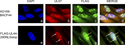
FIG. 4.
Localization of UL57 and FLAG-UL44 proteins in electroporated cells. HFF cells were electroporated with AD169-BACF44 (panels a to d) or BAC-FLAG-UL44-290NLSstop (panels e to h). At 8 days posttransfection, cells were fixed and then stained with antibodies specific for UL57 (Virusys) or FLAG (Sigma), followed by a secondary antibody coupled to fluorophores to detect UL57 (anti-mouse Alexa 594; panels b and f) and FLAG (anti-rabbit Alexa 488; panels c and g) antibodies. DAPI stain was used to counterstain the nucleus (panels a and e). Panels d and h are merged images of the panels in the other columns. White arrows identify punctate UL57 staining. Yellow arrows identify areas of concentration of FLAG-UL44 staining. Magnification: ×1,000.

