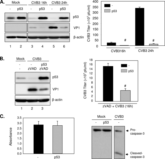
FIG. 5.
Overexpression of p53 inhibits CVB3 infection. (A) HeLa cells were transiently transfected with a plasmid expressing p53 or empty vector pCMV for 24 h, followed by infection with CVB3 (MOI = 1) for 16 or 24 h. Cell lysates were analyzed by Western blotting for protein expression of p53, VP1, and protein loading control β-actin (left panel). Supernatants of infected cells were harvested, and progeny virion titers were measured by a plaque assay (right panel). The results are presented as means ± SD (n = 3). #, P < 0.001 for comparison to vector control cells. (B) HeLa cells were transfected with p53 construct or empty vector for 24 h and then infected with CVB3 for 16 h in the presence of zVAD (50 μM). Western blot analysis (left panel) and a plaque assay (right panel) were performed to examine VP1 expression and virus titers, respectively. The results are shown as means ± SD (n = 3). #, P < 0.001 for comparison with the vector control. (C) HeLa cells were transiently transfected with p53 or empty vector for 48 h. (Left panel) Cell viability was determined by the MTS assay (mean ± SD; n = 3). (Right panel) Western blot analysis for the cleavage of caspase-3. HeLa cells infected with CVB3 were used as a positive control. −, empty-vector control.

