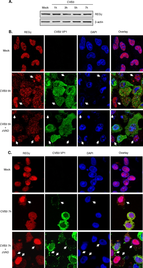
FIG. 7.
CVB3 infection leads to redistribution of REGγ. (A) HeLa cells were either mock infected or infected with CVB3 (MOI = 10) for different times. Western blot analysis was performed for detecting REGγ and β-actin. (B, C) HeLa cells were infected with CVB3 (MOI = 10) for 5 h (B) or 7 h (C) in the presence or absence of zVAD (50 μM). Double-immunocytochemical staining was carried out for examination of the expression and localization of REGγ (red) and viral protein VP1 (green). The nucleus was stained with DAPI (blue). Arrows denote cells without or with low levels of viral protein expression. The yellow staining in the merged image indicates colocalization of these two proteins. It is noteworthy that REGγ is redistributed to the cytoplasm in CVB3-infected cells (green-positive cells) but that REGγ remains in the nuclei of noninfected cells (green-negative cells).

