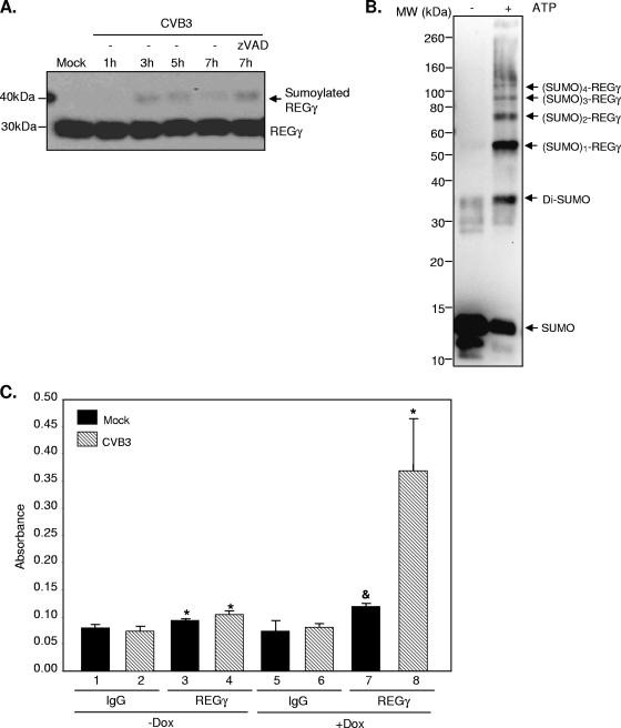
FIG. 8.
CVB3 infection promotes REGγ sumoylation. (A) HeLa cells were infected with CVB3 (MOI = 10) for different times in the presence or absence of zVAD (50 μM). Western blot analysis was performed to detect the expression of REGγ and β-actin. (B) An in vitro sumoylation assay was performed on purified REGγ protein according to the manufacturer's instructions (Biomol). Following in vitro reaction, sumoylated proteins were detected by Western blot analysis using anti-SUMO antibody. (C) HEK293-REGγ stable cells were treated with Dox or without Dox as indicated for 48 h, followed by transient transfection with SUMO-1 for an additional 48 h. After 20 h of mock or CVB3 infection (MOI = 1), cell extracts were harvested for an in vivo sumoylation ELISA as described in Materials and Methods. The data displayed are means ± SD (n = 3). *, P < 0.01 for comparison to mock or CVB3 IgG controls (lane 3 versus lane 1, lane 4 versus lane 2, and lane 8 versus lane 6). &, P < 0.05 for comparison to the mock IgG control (lane 7 versus lane 5).

