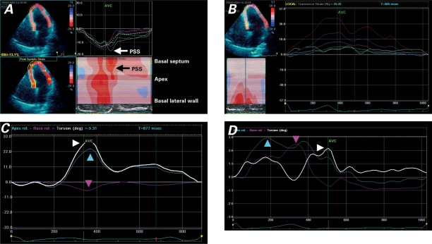
Fig. 2 Speckle-tracking images of the left ventricular septal and lateral walls (multiple views). A) Tracing of longitudinal peak systolic strain shows post-systolic shortening (PSS) in the mid-septal segments. B) Tracing of transverse (radial) strain shows peak systolic strain impairment in the septal segments (yellow, turquoise, and green lines). C) Left ventricular torsion in a healthy patient shows apex rotation (blue arrowhead), base rotation (red arrowhead), and net ventricular torsion (white arrowhead). D) In our patient, net ventricular torsion (white arrowhead) is reduced because of a reduction of apex rotation (blue arrowhead) and a reversal of base rotation (red arrowhead) in early-to-mid-systole; diastolic untwisting is reduced and delayed.
AVC = aortic valve closure

